



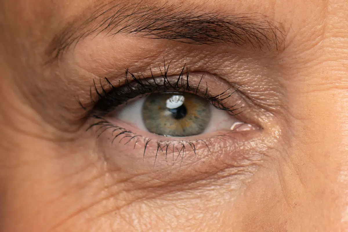
Eyelid ptosis is one of the most difficult clinical conditions to diagnose correctly in oculoplastic surgery, as it is often disguised as what appears to be ageing of the eyelid skin. At Clínica Ramírez & Gálvez, led by Dr. Javier Ramírez and Dr Sonia Gálvez, we often find that a request for cosmetic improvement is actually hiding a dysfunction of the levator muscle. Understanding the nature of eyelid ptosis is essential, because treating it solely as excess skin (dermatochalasis) would lead to an inadequate and poorly functional surgical result for the patient.
Differential diagnosis: ptosis versus dermatochalasis 🔍
It is essential to make a precise technical distinction before any procedure. While dermatochalasis is defined by lax skin that hangs down over the eyelashes, eyelid ptosis is characterised by an abnormally low position of the free margin of the upper eyelid. In severe cases of eyelid ptosis, the eyelid partially covers the pupil, directly compromising the upper field of vision and forcing the patient to compensate by straining the forehead muscle.
During the clinical assessment, we evaluate specific parameters such as levator muscle function and the margin reflex distance (MRD1). This measurement determines whether the problem lies in the “cover” (the skin) or in the “motor” of the eyelid. If we detect eyelid ptosis and go ahead with a standard blepharoplasty without correcting the muscle, the eyelid will continue to look “droopy”, perpetuating the tired or sad appearance the patient wants to get rid of.
Surgical approaches to correcting eyelid ptosis 🎯
Once eyelid ptosis has been confirmed, the surgical strategy is tailored to the strength of the levator muscle and the degree of droop. At our clinics in Lucena and Torre del Mar, we use advanced microsurgery protocols to reattach or shorten the affected muscle. Depending on the patient’s anatomy, eyelid ptosis may be approached via:
- Anterior (external) approach: The natural eyelid crease is used to access the levator muscle. This is the preferred technique when eyelid ptosis is moderate or severe, as it allows a clear view of the levator aponeurosis.
- Posterior (transconjunctival) approach: This is performed from the inner surface of the eyelid, with no external incisions. It is ideal for mild eyelid ptosis or when an extremely quick recovery with no visible marks is the goal.
- CO2 laser technology: We use the laser to make the initial incisions and dissect the tissues, which is crucial in eyelid ptosis surgery to maintain perfect haemostasis and avoid the swelling that would make it harder to fine-tune the eyelid height during the operation.
Medical rigour in the pursuit of symmetry 🏥
Eyelid ptosis surgery is a procedure measured in millimetres. Working under a high-resolution microscope is what allows the eye surgeon to adjust the tension of the muscle with a precision that traditional techniques cannot achieve. At Clínica Ramírez & Gálvez, we understand that eyelid ptosis does not only affect appearance but may also be a sign of other underlying conditions, which is why a thorough pre-operative assessment is non-negotiable.
The stability of the final result in eyelid ptosis depends on in-depth knowledge of orbital anatomy and flawless technical execution. By combining muscle correction with the removal of excess skin, we achieve a more open, vibrant and, above all, functionally healthy look. This 360º approach defines our commitment to excellence in every surgical procedure we perform.
The complexity of the periocular region calls for expert hands that can read every detail of your anatomy. At Clínica Ramírez & Gálvez, we put all our experience and technology at your service so that you regain not only a youthful look but also everyday visual comfort. If you notice that your eyelids are no longer in their original position, we will be delighted to meet you and carry out a technical assessment of your eyes together to determine the most precise approach for your case.